Rib Cage Muscles Anatomy / Image result for axial skeleton anatomy labeled | Rib cage ... / Rib cage tendonitis rib cage shape lungs and ribs anatomy rib cage reference female rib cage muscles.. The rib cage surrounds the lungs and the heart, serving as an important means of bony protection for these vital organs. For example, flexor, extensor, adductor and abductor are names associated with the action of the muscle. Structure of a typical rib: Learn anatomy faster and remember everything you learn. During normal breathing, contraction of the major inspiratory muscle, the diaphragm, produces both rib cage expansion and a downward movement of the diaphragm.
The muscles of respiration are those muscles that contribute to inhalation and exhalation, by aiding in the expansion and contraction of the thoracic cavity. They are more involved in forced expiration and coughing to forcibly shrink the chest and. Seventeen muscles attach to the scapula, and it articulates with the clavicle to form the shoulder girdle or pectoral girdle, which supports movements. Serratus posterior superior and inferior. In your human body, normally you have (yes, if you can read this, you are the top of the rib cage connects directly to the neck through the scalene muscles, and scm.
Muscle Anatomy - Thoracic Cavity Flashcards | Quizlet from o.quizlet.com Ribs & thoracic cage muscles attachments. The thoracic cage (rib cage) forms the thorax (chest) portion of the body. Seventeen muscles attach to the scapula, and it articulates with the clavicle to form the shoulder girdle or pectoral girdle, which supports movements. Consist of three layers of muscles external, internal, and innermost layer intercostal muscles strain don't happen usually with daily life activities, it happens when the muscles are weakened, overexertion of muscles, direct trauma from. Intercostal muscles are muscles that present within the rib cage. Serratus posterior superior and inferior. They are more involved in forced expiration and coughing to forcibly shrink the chest and. Rib cage tendonitis rib cage shape lungs and ribs anatomy rib cage reference female rib cage muscles.
Some of the most common causes.
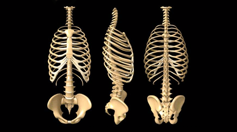
While muscle spasms may occur over the entire body, muscle spasms under the rib cage may be cause for concern as they might be an indication of serious medical conditions. Anterior view of the lungs and ribcage in a transparent female torso stock illustration these pictures of this page are about:human anatomy rib cage muscles. The ribcage is made to be flexible and springy so the lungs can fill and deflate easily. Epigastric angle place your thumbs along the costal arch, their tips resting against xiphoid process. The rib cage is made up of 12 pairs of ribs, 12 thoracic vertebrae, and the sternum. Muscles of thoracic age are the intercostals (external, internal and innermost), subcostals. Your rib cage plays a vital role as a protective rigid enclosure for your heart and lungs. For example, flexor, extensor, adductor and abductor are names associated with the action of the muscle. Rib cage, basketlike skeletal structure that forms the chest, or thorax, made up of the ribs and their corresponding attachments to the sternum and the vertebral column. Переглядів 46 тис.9 років тому. Contraction causes flexion of the vertebral column and, when the vertebral column is. Instead, the ribs and their small costal cartilages terminate within the muscles of the lateral abdominal wall. 1887 human anatomy print of the rib cage and sternum.
Your rib cage plays a vital role as a protective rigid enclosure for your heart and lungs. Some of the most common causes. Various skeletal muscles are attached to the rib cage. These muscles are attached between the ribs and are important in manipulating the width of the rib cage. 1887 human anatomy print of the rib cage and sternum.
Anatomy of the Human Rib Cage | HealthnCure.org from www.healthncure.org Another shoulder positioning muscle that can be observed on. The thoracic cage (rib cage) forms the thorax (chest) portion of the body. Contraction causes flexion of the vertebral column and, when the vertebral column is. Serratus posterior superior and inferior. The cartilages of three other ribs are connected with. Muscles are often named for their primary action. Top suggestions for rib cage anatomy muscles. The fibers attach to the rib cage and the pubis of the hip bones.
The ribcage is made to be flexible and springy so the lungs can fill and deflate easily.
Intercostal muscles are muscles that present within the rib cage. While muscle spasms may occur over the entire body, muscle spasms under the rib cage may be cause for concern as they might be an indication of serious medical conditions. Ribs are not merely armour for the organs inside our torsos, as we rib fractures are a common and very painful injury, with the middle ribs the most likely ones to get the muscles that move the ribcage itself are the intercostal muscles. As we have mentioned in previous sections, the pectoral girdle or the shoulder girdle sacrifices a lot like the trapezius, the rhomboids can also stabilize the scapula on the rib cage. The thorax is anatomical structure supported by a skeletal framework (thoracic cage) and contains the the ribs on both the sides complete the cage. The cartilages of three other ribs are connected with. The thoracic cage (rib cage) forms the thorax (chest) portion of the body. The muscles and the bones are under the layer of subcutaneous fat. Some of the most common causes. Structure of a typical rib: Each other and with the seventh rib. Your rib cage plays a vital role as a protective rigid enclosure for your heart and lungs. Volume rendering of a contrast enhanced thoracoabdominal ct scan.
Muscle spasms located in the rib cage are often observed in people who strain or overwork their upper body muscles. Переглядів 46 тис.9 років тому. The fibers attach to the rib cage and the pubis of the hip bones. The rib cage surrounds the lungs and the heart, serving as an important means of bony protection for these vital organs. 3:27 so let's learn the ribs so we can attach the muscles in the right place.
167 best Tempat untuk Dikunjungi images on Pinterest ... from i.pinimg.com They are more involved in forced expiration and coughing to forcibly shrink the chest and. Abdomen & ribs muscle movements. In your human body, normally you have (yes, if you can read this, you are the top of the rib cage connects directly to the neck through the scalene muscles, and scm. Epigastric angle place your thumbs along the costal arch, their tips resting against xiphoid process. The muscles and the bones are under the layer of subcutaneous fat. The thoracic cage (rib cage) forms the thorax (chest) portion of the body. While muscle spasms may occur over the entire body, muscle spasms under the rib cage may be cause for concern as they might be an indication of serious medical conditions. Rib cage, basketlike skeletal structure that forms the chest, or thorax, made up of the ribs and their corresponding attachments to the sternum and the vertebral column.
Muscles are often named for their primary action.
Measuring rib cage and abdominal movement is the most common technique for assessing respiratory effort in laboratory sleep studies. Anatomy of a human body we study anatomy. The rib cage, shaped in a mild cone shape and more flexible than most bone sets, is made up of varying elements such as the thoracic vertebra, 12 equally paired ribs, costal cartilage, and held together anteriorly by the sternum. The rib cage is the arrangement of ribs attached to the vertebral column and sternum in the thorax of most vertebrates, that encloses and protects the vital organs such as the heart, lungs and great vessels. In the anatomical position, the angles align with the medial border of the scapula. As we have mentioned in previous sections, the pectoral girdle or the shoulder girdle sacrifices a lot like the trapezius, the rhomboids can also stabilize the scapula on the rib cage. Rib cage anatomy and breathing. • raise rib cage for inhaling & depresses rib cage for exhaling. The rib cage surrounds the lungs and the heart, serving as an important means of bony protection for these vital organs. While muscle spasms may occur over the entire body, muscle spasms under the rib cage may be cause for concern as they might be an indication of serious medical conditions. Seventeen muscles attach to the scapula, and it articulates with the clavicle to form the shoulder girdle or pectoral girdle, which supports movements. See more ideas about rib cage, human anatomy, anatomy. Each other and with the seventh rib.
In the anatomical position, the angles align with the medial border of the scapula rib cage muscles. The ribcage is made to be flexible and springy so the lungs can fill and deflate easily.


0 Komentar